The good news is that in most cases instances of neck pain are minor and usually a result of somethingcommonplace: keeping or positioning the head at an awkward angle, “a locked joint” or poor posture while working, reading or watching television. Alternatively, neck pain may be a result of an underlying pathology such as spondylitis, a protruding disc, age-related degenerative disease etc, necessitating diagnosis and treatment by a qualified clinician.
Our heads, which weigh between four and seven kg are balanced on top of our necks with the help of a sophisticated, linked joint system, ligaments and muscles. Accordingly, if these structures are not kept movable, flexible and or strong, especially in the long-term, we are more than likely to suffer from neck problems.
Furthermore, sustained repetitive neck strains and or minor injuries, age-related changes, spine abnormalities, whiplash injuries (including minor repetitive ones) will lead to decreased motion and injury to the supporting structures causing neck pain, irritation (or pinching) of nerves supplying the upper back, chest and arms – leading to something commonly known as “referred pain,” which results in a feeling of pain, tingling and/or numbness radiating down into the upper back, arm, forearm, hand and fingers.

It is estimated that more than 75% of humans will experience low back pain at least once during their lifetimes. The good news is that a large percentage of these patients will recover with minimal therapeutic intervention. The bad news is that the chances of reoccurrence are very high, and thus, it is important that the simplest case be managed properly.
Low back pain is known to arise from trauma (acute or several, i.e., repetitive) such as a fall, a motor vehicle accident, a twisting injury, prolonged poor postures, mental stress, fatigue, disc prolapsed, degenerative disc disease, lost flexibility, decreased muscle strength, reduction in joint motion etc.
Causes such as infection, hormonal problems, visceral dysfunction (such as kidney and gynaecological), broken bones, systemic disease, tumours etc require medical intervention and are beyond the scope of this discussion.
Acute low back pain is defined as pain and dysfunction due to low back or back related leg symptoms (radiating or referred pain, tingling and/or numbness) of less than three months. Chronic low back pain is considered pain that lasts longer than three months. Both types of back pain lead to inflammatory processes in the underlying tissue that produce substances that directly or indirectly cause pain, spasm, stiffness and disability. It is important to understand that it is difficult to determine exactly which tissue(s) is responsible for causing the pain; usually it is a combination of several. It could be muscle(s), ligament(s), disc, joint(s) and/or other connective tissue(s). Interestingly, several of them produce similar symptoms, the common one being pain, and thus diagnosing and pin-pointing the exact cause necessitates systematic assessment and skill. In both cases pain in the low back can be accompanied by pain that radiates down the leg or is limited to the buttock region. Sometimes it may be accompanied by painful spasm in the muscles that support the spine.
X rays are of little help in determining the cause of low back pain, except in

Tendonitis is the inflammation of the tendons; shoulder tendonitis is inflammation of the tendons of the muscles around the shoulder. The signs of inflammation are pain, warmth, redness, tenderness to touch/pressure and loss of function.
Shoulder tendonitis occurs when the rotator-cuff muscles are overloaded, fatigued, traumatized and/or are subjected to age-related degenerative changes. Pinching or impingement of the inflamed tendons that occurs under an overhanging bony structure (called the acromion) is known as the impingement syndrome.
Specifically, impingement happens when a loaded arm is raised repeatedly towards the side over the head or more importantly when the arm is lowered sideways from over the head.
X rays may show a hook or spur that increases the odds that pinching the rotator cuff tendons.
Rotator cuff muscles can also suffer partial and/or complete tears due to an acute, severe fall or fraying due to repeated injuries. The signs and symptoms of tears are pain in the shoulder often radiating down to the middle of the arm, especially when the arm is raised overhead, weakness and in severe cases a complete inability to lift the arm. An MRI is the most common test used for diagnosis. Clearly, complete tears are first treated using arthroscopic surgical techniques and second with movement and therapeutic modalities.
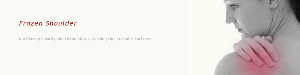
Adhesive capsulitis, or frozen shoulder, is not understood very well. It affects primarily the tissue closest to the joint articular surfaces – the joint capsule. Essentially, pathology progresses with the calcification the capsular tissue up until it is completely calcified. Symptoms include pain and progressive loss of mobility ending with the complete loss of shoulder range of motion.
Causes are not known, but some of the precipitating are as follows: minor traumas, hyperthyroidism, diabetes mellitus, prolonged immobilization of the shoulder, fractures and surgical repairs of structures around the shoulder.
The disease has been characterized as having a freezing, frozen and thawing stage and has been said to be self-limiting having a six month to a two year cycle.

Patients who suffer from golfer’s elbow are often involved with racquet sports or golf. As with tennis elbow it may be as a result of overuse of the medial forearm muscles (common flexors of the wrist and fingers) and/or traumatizing the elbow by hitting several golf shots using a “poor” swing technique or repeatedly hitting the turf (especially hard ones).
Patients usually complain of pain at the inner aspect of the elbow and the inability to use the arm and hand. Symptoms can be usually reproduced with resisted finger and wrist flexion.
The condition is called Golfer’s Elbow because in making a golf swing this tendon is stressed, especially if a non-overlapping (baseball style) grip is used; many people, however, who develop the condition have never handled a golf club. It is also sometimes called Pitcher’s Elbow due to the same tendon being stressed by the throwing of objects such as a baseball, but this usage is much less frequent.
This condition is characterized by pain along the lateral aspect of the elbow and the inability to use or grasp objects with the affected hand. Interestingly this disorder, contrary to common perception afflicts only five percent of people who play tennis.
It is important to point out that pain in and around the elbow could also be due to referred pain from the cervical spine and or irritation of nerves supplying the elbow any way along the course of the nerve. Thus, it is important to differentially diagnose the exact cause of elbow pain before planning its management.
As with golfer’s elbow, patients with tennis elbow have extreme difficulty using their affected hand to perform simple day-to-day activities such as opening doors, picking-up objects, grasping a fork or a spoon.

Carpal tunnel syndrome is a result of the compression of a nerve (median) due to inflammation within the carpal tunnel at the level of the wrist joint. It is characterized by pain, tingling, and in severe cases, numbness in the thumb, index, middle and half of the ring finger.
It is typically caused by repetitive tasks involving the hand and the wrist, for example typing with the wrists resting on hard surfaces with the wrist joints cocked (in extension). It is often worse at night or driving and in very severe cases can lead to a weak grip strength and decreased coordination. If left untreated it can progressively lead to atrophy of the thumb and finger muscles. Women are afflicted frequently more than men.
The main symptom of CTS is intermittent numbness of the thumb, index, long and radial half of the ring finger. The numbness usually occurs at night because we tend to sleep with our wrists flexed and is relieved by wearing a wrist splint that prevents flexion.

Strains and sprains are injuries that we sustain on a daily basis while engaged in day-to-day, recreational, and competitive activities.
Strains refer to injuries that affect muscles, specifically the minor tearing of muscle fibres, which forms the basic unit that is responsible for contraction of the muscle. These injuries commonly occur when the muscle performs repeated strong contraction when it is either overloaded or fatigued and in a weakened state.
Strains can be classified as:
Mild (grade I) – mild disruption of muscle fibres manifested by mild tenderness and slight pain with active use or stretch
Moderate (grade II) – indicates moderate damage to the fibres and is manifested by tenderness, some amount of distal discolouration (or bruising) caused by damage to the small blood vessels, and some degree of pain and tenderness in the damaged muscle.
Severe (grade III) – is due to the partial or complete tearing of muscle from its associated tendon. It is manifested by severe pain, complete loss of power, swelling, bruising and a palpable “dip” or “indentation” where the muscle has been damaged. If there is a complete tear of the muscle – the only solution is surgical intervention.

Sprains are tears of the non-contractile supporting structures of the joints, ligaments and capsule. These structures consist of limited elastic collagen tissue that intimately attaches one articulating bone to another. Further, damage to the capsule leads to loss of nutrients and lubrication fluid (synovial fluid) of a joint, leading to serious joint damage. Similar to strains, sprains are likewise categorised as follows:
Grade I – involves the tearing of a minimal number of ligament fibres. There may be pain, swelling, and some loss of motoric function.
Grade II – involves a moderate or partial ligament tear. There is moderate to severe pain, swelling, tenderness and loss of joint function. Added to this is the loss of functional ability such as walking.
Grade III – involves the complete tearing of the ligament or joint capsule, which is sometimes accompanied by avulsion fractures. It is associated with severe pain, swelling and loss of function and most often requires immobilization and/or surgical intervention.

Osteoarthritis is the most common form of arthritis that afflicts joints and literally means inflammation of a joint. OA is described by inflammation and degeneration of the cartilage (hyaline) that protects the articular surfaces of the joint, usually the knee joint. It is important to note that this cartilage provides a cushion and thus plays an important role in dissipating load forces and also protecting the underlying bone. If damaged, it, over a period of time, leads to exposing the bone and the growth of bone spurs (osteophytes). Accordingly, it leads to a viscous cycle of pain <-> inflammation <-> damage.
It is a fact that most adults over a period of their life times will develop OA. Research has identified the following factors that predispose to this disabling disease:
- Obesity/Weight – the more the obese or the heavier a person the more likely he will experience OA
- Trauma – joints that have experienced trauma, particularly repetitive, are more likely to develop OA
- Infection or other pathologies can lead to joint damage and the development of arthritisOA
Subjects suffering from OA complain primarily of pain, joint stiffness after long periods of inactivity, “catching” or “locking” of joints, and a feeling of grinding. The common signs associated with it are pain, tenderness, warmth, loss of range of motion, decreased muscle strength and functional disability such as problems walking, getting up, running etc. Severe cases of OA will need to resort to joint replacements

This disorder is characterized by chronic pain and tender points and/or nodules within muscle tissue across large areas of the body. It is said to be part of an autoimmune spectrum of disorders. The tender nodules, which hurt when they are pressed, are found in specific places – neck, shoulders, hips, back and legs and patients present with a broad spectrum of associated complaints:
- Stiffness
- Fatigue
- Sleep disturbance
- Headache
- Depression
- Impaired muscular performance

This refers to the loss of bladder and bowel control, resulting in involuntary leakage of urine or faeces primarily due to weakness of the pelvic floor musculature (pelvic muscles). Weakness of the muscles leads to a loss of support and control of the bladder and bowels and thus patients are unable to voluntarily open or close the openings, to leakage of content. This leakage becomes even more of a problem with physical stress (coughing, sneezing, and laughing) or when the bladder or bowel is full. In addition, muscular dysfunction leads to a problem of urgency, i.e., involuntary emptying whenever the sensation of urge is felt.
Both conditions are commonly seen after surgical procedures on organs in the pelvic cavity, pregnancy and child birth. These conditions respond successfully to remediation using several rehabilitation techniques such as functional electrical stimulation, biofeedback and others.

A stroke, or cerebrovascular accident (CVA), is the rapid loss of brain function due to disturbance in the blood supply to the brain and the resultant damage to the nervous tissue. As a result of this damage there may be an inability to move one or more limbs on one side of the body, inability to understand or formulate speech and/or deficits in vision.
Stroke rehabilitation is the process by which those with disabling strokes undergo treatment to help them return to normal life as much as possible by regaining and relearning the skills of everyday living. It also aims to help the patients understand and adapt to difficulties, prevent secondary complications and educate family members to play a supporting role.
Physiotherapy focuses on joint range of motion and strengthening exercises and the re-learning of functional tasks of such as bed mobility, transferring, walking and other gross motor functions. Therapists also work with patients to improve body awareness, the use of the hemiplegic side, faulty balance and foot drop.
Rehabilitation focuses on facilitating the production of strong movements using normal patterns during the performance of day-to-day functional tasks. Emphasis is often on functional tasks and the patient’s individual goals. Through continuous practice the patient relearns to use and adapt the hemiplegic limb during functional activities to create lasting permanent changes and independence in day-to-day activities.

Physiotherapists play a very important role in the rehabilitation of patients who have sustained fractures. They are usually referred for therapy after a period of immobilisation – usually 6 to 12 weeks.
Physiotherapy helps accelerate the healing process, reduce pain and swelling and improve range of motion to get the patients back to their normal physical self. Physiotherapy is most often incorporated into a patient’s healing routine after sufficient healing has taken place and the bone is properly aligned. It is purported to strengthen the bone and the muscle tissue surrounding the bone in order to assist the patient regain full range of motion and independent mobility and function.
Physiotherapy uses a variety of modalities and exercises to help in its rehabilitation. The modalities used but not limited to are, Ultrasound, TENS, Hydrotherapy, Paraffin wax bath or Electrical stimulation.
Exercises will help to develop the muscles and improve strength and mobility in and around the affected area. Different exercises such as active, passive, resisted, concentric/eccentric etc are used in combination with modalities to improve range of motion, strength, flexibility in the affected joint and muscle – with the goal to achieve complete functional independence.
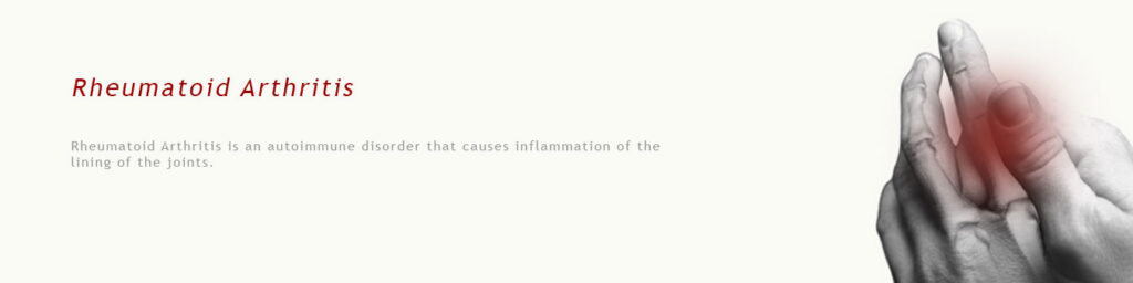
Rheumatoid Arthritis is an autoimmune disorder that causes inflammation of the lining of the joints. The body tissue is mistakenly attacked by its own immune system. It may also affect the skin, eyes, lungs, heart, blood or nerves. Women are affected three to five times more often than men.
Joints become swollen, tender warm and stiff, all of which limit their movement. With time multiple joints are affected. Most commonly involved are the small joints of the hands, feet and cervical spine, but larger joints like the shoulder and knee can also be involved.
Patients with RA present with a combination of the following symptoms:
- Morning joint stiffness
- Swelling/fluid around several joints of the body
- Reduced range of motion
- Reduced muscle strength
- Debility and generalized weakness
Onset is uncommon under the age of 15 and from then on the incidence rises with age until the age of 80.
The goals of treatment are to minimize symptoms such as pain and swelling, to prevent bone deformity, and to maintain day-to-day functioning. Primarily treatment includes a combination of exercises to strengthen supporting muscles around the joints. Regular supervised exercises of stiff and painful joints reduces the overall pain in patients with RA. In addition, exercises keep bones and muscles strong and help reverse joint stiffness.
Modalities such as heat, Paraffin wax bath, Ultrasound, TENS, Interferential Therapy etc are used to alleviate pain and joint stiffness.

Shin splints (also known as Medial tibial stress syndrome or tibial periostitis) is a common injury that affects athletes who engage in running sports or physical activity. This condition is characterized by pain in the lower part of the leg just above the ankle and is caused by repeated trauma to the tissue contained in the anterior compartment of the leg and the tissue surrounding the tibial bone. Ignoring these injuries may result in a more serious condition such as a stress fracture of the bones.
Shin splints or MTSS can usually be attributed to overloading the muscles of the lower extremities or to biomechanical discrepancies. Though the exact cause of this condition is not known, it is usually precipitated by a change in running routine, change of footwear and/or a change in running surface. In addition, a combination of weak musculature made to overwork plus biomechanical insufficiency underlies most cases of shin splints.
Thus, muscle imbalance, including weak core muscles, inflexibility and tightness in the musculature of the lower extremities may contribute to MTSS.
A typical clinical presentation of this condition involves pain, palpable tenderness, and possible swelling.
MTSS is initially treated with rest, ice to reduce inflammation, NSAIDs, and Physiotherapy.
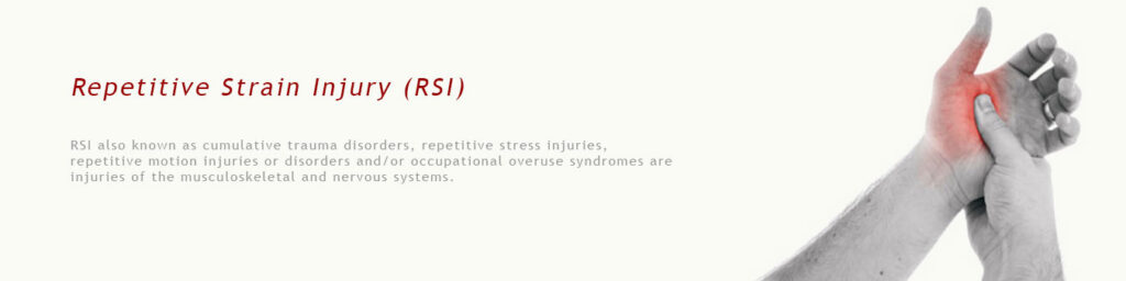
RSI also known as cumulative trauma disorders, repetitive stress injuries, repetitive motion injuries or disorders and/or occupational overuse syndromes are injuries of the musculoskeletal and nervous systems that are precipitated by repetitive tasks, forceful exertions, vibrations, mechanical compressions or sustained and/or awkward positions.
Patients generally present with activity-related limb pain, tingling, numbness, stiffness and/or fatigue which increases as the activity is sustained.
Disorders that fall under this category are – Shin splints, Carpal/Tarsal Tunnel Syndromes, Golfer’s/Tennis elbow and even non-specific disorders popularly referred to as Blackberry thumb, Ipod finger, gamer’s thumb, trigger finger.
Nerve Conduction Velocity tests, X-rays and MRIs are some of the imaging techniques used to provide a confirmatory diagnosis.
Management of these disorders is customized to the individual patient. Treatment most often consists of a detailed assessment followed by a combination of modalities and an individualized exercise program.
Generalised exercise has been shown to decrease the risk of developing RSI.